causes of joint pain
lots of physical activity
Age-related changes in the musculoskeletal system
Pregnant
- vitamin and mineral deficiencies. The greatest role played by calcium and vitamin D deficiencies is in the development of osteomalacia. A characteristic feature of the symptom presentation is not only joint pain, but also bone pain, fatigue, the occurrence of hypocalcemia and other symptoms of vitamin D deficiency - dental caries, brittle nails, muscle weakness, muscle pain and the frequent occurrence of ARVI.
- significant weight gain. Joint discomfort is more common in pregnant women who have gained a lot of weight or are obese. Pain is felt at the end of the day and eventually in the middle of the day in the hip joints, knees, ankles, where the cartilage is exposed to loads several times higher than allowed. To alleviate this condition, women will deliberately limit physical activity, causing weight gain to occur more quickly.
- Softening of cartilage and ligaments. About half of pregnant women experience pelvic joint discomfort due to the effects of the hormone relaxin. In most cases, the nature of the discomfort is pain in the pubic area and hip joints. In the pathological process of developing syndesmitis, the soreness is replaced by pain, which is aggravated by pressure on the uterus during sexual intercourse and when trying to spread the legs. Pain in the pubic area is a serious reason to see your ob-gyn.
- carpal tunnel syndrome. Nearly 20% of pregnant women develop a special manifestation, the so-called tunnel syndrome, at 2-3 months of pregnancy. The disease is caused by swelling of the soft tissues of the hand and compression of the carpal tunnel of the nerves that carry them to the fingers. In addition to pain in the facet joints of the hands, patients complain of numbness, tingling, and a crawling sensation in the skin. This condition improves as the arm position is raised.
obesity
acute infection

Collagenopathy
- Rheumatism. The symptoms are "unstable": aches and pains in the large joints of the arms and legs (elbows, shoulders, hips, knees, ankles) in sequence. Swelling of the affected area. Joint discomfort is often preceded by a sore throat. With treatment, joint changes are reversible.
- Rheumatoid Arthritis. Unpleasant feelings often appear after 40 years. Typical pain in the facet joints of the hands and feet, with significant swelling and morning stiffness. In the future, pain and curvature of the joints will become more prominent.
- systemic scleroderma. It is characterized by localized changes in pain sensation and stiffness in the hands, elbows, and knees in the morning. The pain is usually symmetrical. The swelling is short-lived. Due to hardening of the skin, joint movement is limited, tendons are damaged, and friction occurs during movement.
Osteoarthritis
Metabolic disorders
- osteoporosis. When calcium is washed away from bone tissue, the joint surfaces of the bones become fragile and the cartilage becomes thinner, accompanied by pain. The pain syndrome progresses from mild pain to severe joint pain, accompanied by skeletal discomfort and muscle weakness. The joints that bear the greatest load are most commonly affected - the hips and knees; the shoulders, elbows and ankles are less commonly affected.
- gout. Mild pain in the big toe is already a cause for concern during the preclinical stages of the gout process. Pain and discomfort may occur in the knees, elbows, wrists, and fingers. The accumulation of urate in the joint space causes the disease to manifest rapidly, from aches and pains to acute pain that does not subside within several hours. The affected joint is hot to the touch. Red skin and limited movement.
neoplastic disease
joint damage
chronic infectious process
Complications of drug therapy
- antibiotic: Penicillins, fluoroquinolones.
- sedative: Phenazepam, diazepam, lorazepam, etc.
- contraceptives: Combined oral contraceptive (COC).
Rare causes
- Respiratory system inflammation: Pneumonia, bronchitis, tracheitis.
- intestinal pathology: Non-specific ulcerative colitis, Crohn's disease.
- skin disease: Psoriasis.
- Endocrine disorders: Diabetes mellitus, diffuse toxic goiter, hypothyroidism, Itsenko-Cushing disease.
- autoimmune process: Hashimoto's thyroiditis, vasculitis.
- fascia injury: Necrotizing fasciitis during recovery.
- congenital bone and joint defects.
opinion poll
- laboratory blood test. Evaluation of white blood cell count and ESR levels is required to rule out infectious, inflammatory, and oncohematological processes. In systemic diseases, it is important to measure total protein content, the ratio of protein components in the blood, specific acute phase proteins, markers of rheumatoid arthritis and other inflammations. Tests of vitamins, electrolytes (especially calcium), and uric acid concentrations can help diagnose metabolic disorders.
- Bacteriological examination. If joint and general pain may be contagious, a bacterial culture may be required. Urine, feces, sputum, and urogenital secretions were collected for study. In order to select an antimicrobial treatment regimen, susceptibility to antibiotics needs to be determined. In suspicious cases, microscopy and culture are supplemented by serological reactions (RIF, ELISA, PCR).
- Joint Ultrasound. It is often used to clearly localize pain and suspect the presence of rheumatic disease. Ultrasound examination of joints allows us to examine their structure, identify destruction of cartilage and bone, preclinical inflammatory changes, and study the condition of the soft tissues surrounding the joint. The advantages of this method are accessibility, non-invasiveness, and high information content.
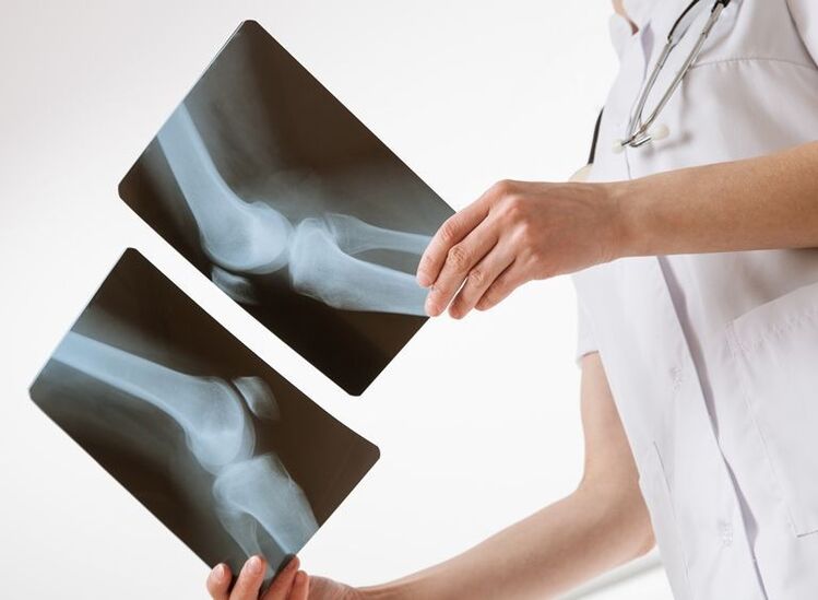
- X-ray technology. Changes in joint space width, soft tissue sclerosis, the presence of calcifications, osteophytes, and articular surface erosion can be detected during joint radiography. To increase the efficiency of diagnosis, special techniques are used - contrast arthrography, pulmonary angiography. In the initial stages of pathology, tomography (MRI, joint CT) is considered more indicative. Bone density can be easily assessed using densitometry.
- Invasive inspection techniques. In some cases, to determine the cause of joint pain, an aspiration may be performed to take a biopsy of the cartilage, synovial lining, and tophi. Morphometric analysis of biopsy specimens and examination of synovial fluid reflect the nature of the pathological process occurring in the joint. During arthroscopy and tissue biopsy, material collection and visual inspection of the joint space can be conveniently performed simultaneously.

treat
Pre-diagnosis help
Conservative treatment
- Antibacterial agents. The basic treatment of infection is based on the prescription of antibiotics sensitive to the pathogen. In severe cases, broad-spectrum drugs are used until the susceptibility of the microorganism is determined.
- NSAIDs. They reduce the production of inflammatory mediators and thus suppress the inflammatory process in the joints. By affecting central pain receptors, they can reduce the level of joint discomfort. Available in the form of tablets, ointments, and gels.
- corticosteroids. They have strong anti-inflammatory effects. Hormone therapy is the basis for the treatment of systemic collagenopathies. In severe and resistant disease, corticosteroid drugs are combined with immunosuppressants to enhance efficacy.
- chondroprotectant. They serve as substrates for the synthesis of proteoglycans, which in sufficient amounts increase the elasticity of articular cartilage. Nourish cartilage tissue and restore its damaged structure. Intra-articular administration is possible.
- xanthine oxidase inhibitor. Used as an anti-gout medicine. They block key enzymes required for the synthesis of uric acid, thereby reducing its concentration in the body and promoting the dissolution of existing urate deposits.
- vitamin mineral complex. Recommended for the treatment of joint pain caused by metabolic disorders. The most commonly used medications contain calcium and vitamin D. They are also an element in the comprehensive treatment of inflammatory and metabolic diseases.
- Chemotherapy drugs. They are the basis of most treatment options for various types of neoplastic hematological pathology. Depending on the clinical variation and severity of the new process, they are combined with radiotherapy and surgical intervention.


